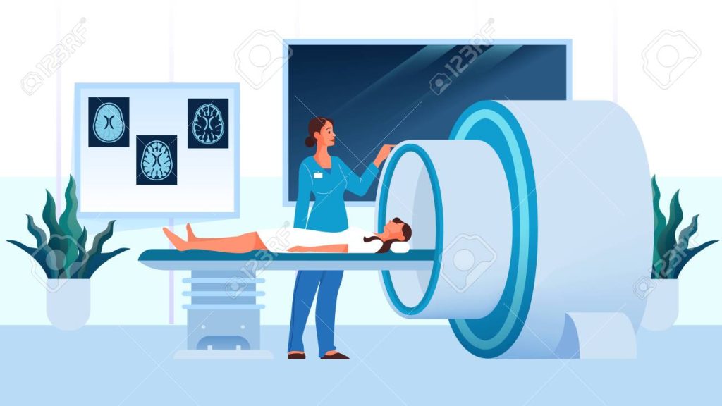
The full form of MRI is Magnetic Resonance Imaging. MRI is also known as NMRI (Nuclear Magnetic Resonance Imaging) or MRT (Magnetic Resonance Tomography). MRI is a diagnostic imaging technique used in radiology to display more information about the internal body structures clearer than X-Ray. It can be used to create representations of the anatomy and physiological functions of the body in both illness and health.

History of MRI
MRI’s captivating history:
- NMR Discovery (1938): Isidor Rabi’s discovery of nuclear magnetic resonance (NMR) laid MRI’s foundation.
- Early NMR Exploration (1940s-1950s): Scientists explored NMR, paving the way for medical imaging.
- First MRI Prototype (1970s): Paul Lauterbur and Raymond Damadian independently proposed MRI for medical imaging. Lauterbur introduced spatial encoding, while Damadian built the first MRI prototype.
- Clinical MRI Scanners (1970s-1980s): Clinical MRI scanners emerged, though early machines were large and time-consuming.
- Advancements (1980s-Present): MRI evolved with high-field magnets, faster sequences, fMRI for brain activity, DWI, and MRA for diagnostics.
- Clinical Impact: MRI revolutionized diagnosis, detecting various conditions non-invasively.
- Ongoing Innovation: Research in ultra-high-field imaging, molecular MRI, and advanced contrast agents promises enhanced diagnostics.
How MRI Works
MRI, or Magnetic Resonance Imaging, is a medical imaging technique that reveals detailed internal structures of the body. Here’s a simplified breakdown of how it works:
- Magnetic Fields: Patients in an MRI machine are exposed to a strong magnetic field that aligns the protons (hydrogen nuclei) in their bodies.
- RF Pulses: Short bursts of radiofrequency waves (RF pulses) are directed at the body’s target area, temporarily disrupting proton alignment.
- Relaxation Process: When RF pulses stop, protons return to alignment and emit RF signals as they do so.
- Signal Detection: Specialized coils within the MRI machine detect these RF signals, which vary based on tissue type.
- Image Creation: A computer processes these signals, generating cross-sectional images of the body, creating a 3D image.
- Contrast Agents: Sometimes, contrast agents are used to enhance specific areas, making them appear brighter on MRI images.
- Medical Interpretation: Radiologists analyze MRI images to diagnose various conditions, leveraging the contrast and structural details.
Types of MRI Scans
MRI scans are valuable for diagnosing various conditions in special parts of the frame:
- Brain MRI: Images the mind and spinal wire, assisting in the diagnosis of tumors, strokes, infections, and a couple of sclerosis.
- Spinal Cord MRI: Focuses at the spinal cord and surrounding structures, helping in diagnosing spinal stenosis, herniated discs, and tumors.
- Chest MRI: Captures images of the chest, along with lungs and coronary heart, to diagnose situations like lung cancer, coronary heart ailment, and pneumonia.
- Abdominal MRI: Examines stomach organs just like the liver, pancreas, and kidneys, aiding in the diagnosis of liver cancer, pancreatitis, and kidney stones.
- Pelvic MRI: Targets pelvic organs like the uterus, ovaries, and prostate gland, supporting in diagnosing conditions such as endometriosis, ovarian most cancers, and prostate most cancers.
MRI Equipment and Machines
MRI machines are vital tools in modern medicine, offering non-invasive and detailed body imaging. They come in various types:
- Closed MRI: Traditional, powerful, but potentially claustrophobic due to the enclosed tunnel.
- Open MRI: Patient-friendly with a more open design, though slightly lower magnetic field strength.
- High-Field MRI: Strong magnetic fields (1.5T to 3.0T) for excellent image quality, suitable for various applications.
- Ultra-High-Field MRI: Extremely strong magnetic fields (7T to 9.4T+) for advanced research and specialized clinical uses.
- Portable MRI: Compact and mobile, ideal for point-of-care imaging in emergencies or remote areas.
- Dedicated Extremity MRI: Focuses on specific body parts, like extremities, providing high-resolution images for orthopedic purposes.
- Open-Bore MRI: Blends openness with high image quality, enhancing patient comfort.
Conclusion
In conclusion, Magnetic Resonance Imaging (MRI) stands as a remarkable achievement in the field of medical diagnostics. This non-invasive and radiation-free imaging technique has revolutionized the way we visualize and understand the human body’s internal structures. Its versatility, ranging from brain scans to extremity imaging, allows for the comprehensive assessment of numerous medical conditions.
FAQs About MRI
Yes, MRI is considered safe because it doesn’t use ionizing radiation like X-rays or CT scans. However, certain conditions or metallic implants may pose risks, so it’s important to inform your healthcare provider of any relevant medical history.
The duration of an MRI scan varies depending on the type and body part being imaged. It can range from 15 minutes to over an hour.
No, MRI itself is not painful. However, some patients may experience discomfort from lying still for an extended period or due to the noise produced by the machine.
Contrast agents containing gadolinium may be used to enhance specific areas of the body. Your healthcare provider will determine if contrast is necessary for your scan.


